
The initiative aims to support clinical diagnosis and scientific research by utilizing imaging data in combination with other health markers, while simultaneously establishing a national brain scan repository that will serve as a centralized resource for advancing neuroscience and medical practice in the Philippines.
Clinical and Neuropsychological Assessment
In our team, we conduct comprehensive behavioral and cognitive assessments to gain a full picture of each participant’s memory, thinking skills, mood, daily functioning, and overall health. We start by collecting detailed medical history, current medications, and key physical measurements, then administer a wide range of standardized tests—including the Clinical Dementia Rating, Montreal Cognitive Assessment–Philippine version (MoCA-P), Geriatric Depression Scale, and Instrumental Activities of Daily Living (IADL) scale. We also evaluate quality of life (EQ-VAS), Alzheimer’s-related cognitive changes (ADAS-Cog), and executive functions through tasks such as verbal fluency, Trail Making, digit symbol substitution, and number cancellation. To complement these evaluations, we interview caregivers using informant-based tools like Alzheimer's Disease 8 (AD8), Disability Assessment for Dementia (DAD), and Neuropsychiatric Inventory–Questionnaire (NPI-Q). Each session, typically lasting about 90 minutes with breaks as needed, reflects our department’s commitment to thorough, culturally attuned assessment of cognitive health and functional abilities.
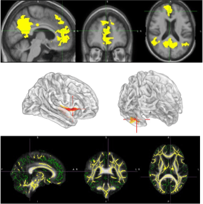
fMRI
Functional MRI (fMRI) is a high-spatial-resolution non-invasive neuroimaging technique used to visualize brain activity by detecting Blood Oxygenation Level Dependent (BOLD) signals. These signals originate from dynamic changes within tissue vasculature due to alterations in blood flow, volume, and oxygenation, reflecting neural activity. There are two main types: task-based fMRI (tb-fMRI), which measures activation during specific tasks, and resting-state fMRI (rs-fMRI), which identifies intrinsic connectivity networks (ICNs) or resting-state networks (RSNs) without a task. It helps to identify areas of altered brain activation and functional connectivity, providing insights into a variety of neurological and neuropsychological conditions.
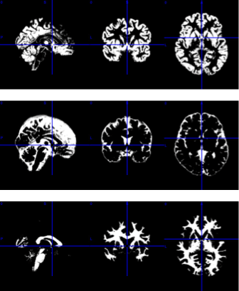
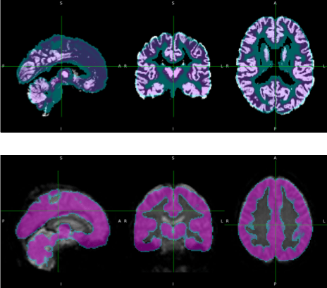
STRUCTURAL MRI
Structural MRI is a non-invasive imaging technique that provides detailed visualization of brain anatomy and detect structural lesions or abnormalities. It characterizes brain structure, focusing on detecting associated structural abnormalities like tumors, stroke, or degenerative changes, but does not directly assess functional dynamics. It serves as an anatomical reference for other functional and advanced techniques like fMRI and DTI. This is crucial for diagnosing neurological disorders, tracking disease progression, and understanding the impact of various conditions on brain structure.
DTI
Diffusion Tensor Imaging (DTI) is an advanced structural MRI technique that provides detailed visualization of brain anatomy, particularly focusing on white matter integrity. It can detect structural abnormalities and physical cortical disconnections by analyzing the anisotropic diffusion of water molecules. This technique helps define the anatomy of normal white matter tracts and identifies changes caused by various neuropathological processes. DTI is an ideal tool for investigating white matter and its participation in cognitive and emotional functions. DTI helps understand the brain's structural connectivity that underpins functional networks.
F-fluorodeoxyglucose (FDG) brain positron emission tomography (PET)
"F-fluorodeoxyglucose (FDG) brain positron emission tomography (PET) is used to map glucose metabolism throughout the brain. FDG is a radiotracer that functions as a glucose analogue. The brain – as an obligate glucose user – preferentially accumulates FDG in its tissues, where patterns of FDG uptake reflect neuronal and synaptic activity. The radiotracer emits positrons and produces annihilation photons, which are detected by the PET scanner to generate metabolic images. An integrated computed tomography (CT) scanner provides anatomic data for improved morphologic correlation. PET/CT abnormalities in the brain typically represent underlying pathologies, such as epileptogenic foci, tumors, vascular lesions, and neurodegenerative diseases."
Image processing and analyses are predominantly conducted using MATLAB and Python. External plugins, including SPM25 and CAT12, facilitate the preprocessing of sMRI, fMRI, and PET images. The preprocessing pipeline encompasses segmentation, slice timing correction, motion correction, coregistration, normalization, and smoothing. FSL, a Python-based library, assists in the preprocessing and analysis of dMRI and fMRI. Additionally, MRIcron is employed for image visualization and comparison.
Statistical analyses are employed to compare group differences. Structural differences are assessed using voxel-based morphometry, surface-based morphometry, and deformation-based morphometry. Functional differences are measured through seed-based correlation analysis and independent component analysis. Diffusion tract measurements are compared via tract-based spatial statistics and voxelwise statistics. PET images are analyzed using standard uptake value measurements.
In this stage, images are processed for motion correction, denoising, smoothing, modulation, and alignment between modalities

Imaging Data and Analysis
Clinical and Neuropsychological Assessment
Structural MRI
Functional MRI
DTI
FDG-PET
Neuromap-PH
info@neuromap.ph